2026-09-03
Surgical Tips for Aesthetic Lower Lid Blepharoplasty: Prevention of Round Eye I
PRS Global Open, 2023년에 실린 저널Surgical Tips for Aesthetic Lower Lid Blepharoplasty: Prevention of Round EyeKlinger, Marco MD*; Vinci, Valeriano MD†,‡; Romeo, Maria Angela MD†; Battistini, Andrea MD*;
PRS Global Open, 2023년에 실린 저널
Surgical Tips for Aesthetic Lower Lid Blepharoplasty: Prevention of Round Eye
Klinger, Marco MD*; Vinci, Valeriano MD†,‡; Romeo, Maria Angela MD†; Battistini, Andrea MD*; Klinger, Francesco MD§; Bandi, Valeria MD*; Maione, Luca MD*; Vinciguerra, Paolo MD†,¶; Di Maria, Alessandra MD†,¶
INTRODUCTION
Lower eyelids are extremely predisposed to the effects of aging, such as skin laxity, loss of ligamentous support, atonia, and lower orbicularis-oculi muscle ptosis, as well as thinning of the orbital septum, which leads to orbital-fat prolapse.1,2
하안검은 aging에 매우 취약한데, 특히 skin laxity, loss of ligamentous support, atonia, and lower orbicularis-oculi muscle ptosis 등의 aging이고, orbital septum이 얇아지면서 orbital fat이 튀어나오게 된다.
an overall evaluation of different orbital structures is paramount to optimize lower lid rejuvenation.
Lower eyelid malposition..
scleral show vs ectropion!!
|
항목 |
Scleral Show |
Ectropion |
|
정의 |
아래 눈꺼풀 아래로 **공막(sclera)**이 노출되어 있는 상태 |
아래 눈꺼풀이 바깥쪽으로 돌출되어 결막이 외부로 노출된 상태 |
|
형태 |
하안검이 약간 아래로 처져 있어 흰자위가 많이 보임 |
하안검이 완전히 뒤집혀 속눈썹과 결막이 외부로 노출됨 |
|
중증도 |
경미하거나 해부학적 변이일 수도 있음 |
기능적인 문제를 일으킬 수 있는 병리적인 상태 |
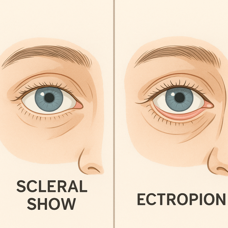
🔹 Scleral show는 아래 눈꺼풀이 살짝 내려가서 흰자위가 보여서 미용적으로 보기 싫은 상태
🔹 Ectropion은 아래 눈꺼풀이 완전히 말려 올라가서 기능에 문제를 주는 병적 상태
MATERIALS AND METHODS
Clinical Assessment and Patient Selection
Between January 2016 and January 2020,
bilateral lower lid blepharoplasty on 280 patients at Humanitas Research Hospital (Rozzano, Milano, Italy)
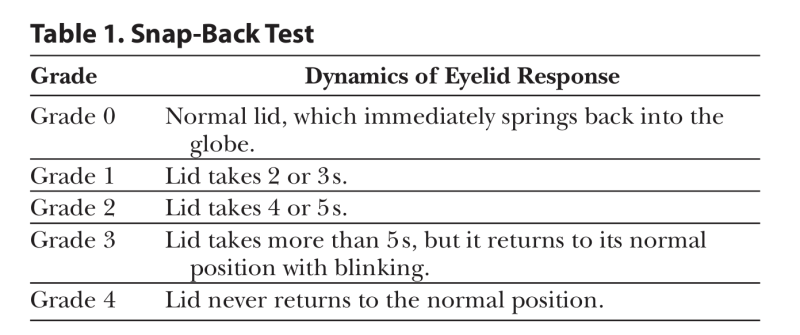
All patients were evaluated preoperatively in terms of ocular surface parameters: (a) lower eyelid retraction using “scleral show,” defined as the visible portion of the sclera between the upper rim of the lower eyelid and the inferior limbus; (b) lower eyelid laxity using the snap-back test (Table 1), the distraction test, and the assessment of eyelid margin malposition relative to the globe9; (c) amount of skin excess; (d) asymmetry; and (e) presence/ absence of orbital fat herniation (7.0.1. Adobe Photoshop Inc, California, was used to measure the distance).
Routine complete blood count and ECG were per formed in all patients. Preoperative standard photographs were collected, with the camera at a fixed 1-meter distance. Blinded-fashion preoperative and 10-month postoperative photograph analysis was performed by one independent plastic surgeon and one oculoplastic surgeon who did not execute the procedures to estimate both functional and aesthetic outcomes. Hence, postoperative photographs were scored from 1 (unsatisfactory outcome) to 5 (opti mal outcome).
Finally, at 10-month follow-up visit, a VAS from 0 to 10 was administered to all patients to assess patient sat isfaction in terms of lower lid appearance and symmetry compared to baseline. The VAS was shown to be less vul nerable to bias from confounding factors compared to other scales.10
Takeaways Question: How to prevent surgical complications in lower eyelid blepharoplasty? Findings: We highlight novel insights into a surgical technique for lower eyelid reconstructive surgery based on our 20-year experience in this specific field, which avoids the use of muscle flaps, thus preventing tarsal liga ment dystopia, limiting thermal spread, and preserving orbicularis-muscle innervation. We discuss clinical mani festations, such as epiphora, dry eye, blurred vision, eye discomfort, and photophobia, and their relation to lower lid displacement.
Meaning: Our technique helped us in preventing surgical complications, specifically lateral canthal dystopia, which ranges from round eye to ectropion. This improves post operative outcomes, revealing high patient and surgeon satisfaction over time.
A longstanding and satisfying experience with this technique let our group develop peculiar shrewdness crucial in the prevention of round eye. Particularly, our expertise focuses on the following strategical aspects:
1. The correction of horizontal skin laxity trough fixation of the lateral portion of the orbicularis oculi. Two possible surgical approaches, with or without the use of muscle flaps, can be used:
Reidy-Adamson technique: a triangular flap is harvested in the upper lateral portion of the lower orbicularis-oculi muscle. This is passed beneath a superolateral canthal tunnel and then fixed at the periosteum of the upper lateral orbital rim (above the Whitnall tubercle) to obtain optimal lower eyelid contour.
Our alternative technique, which does not imply the use of a muscle flap: during the skin excision, the orbicularis-oculi muscle is removed in a complementary fashion. Skin stitches are positioned to grasp skin-muscle-skin in an oblique fashion. This lateral vector puts into tension the muscle, compensating the horizontal skin laxity. As this approach has appeared to be very effective in a relatively short operative time, it has been used as our routine technique for more than 20 years now.
2. Sparing of tarsal ligaments, through a transcutaneous lower lid incision made 1 mm below the eyelashes line, directed to the prevention of dystopia following the tarsal ligament healing process and subsequent ectropion. In fact, misposition of tarsal ligament may lead to palpebral and bulbar conjunctiva detachment, altering the lateral final aesthetic profile of the patient.
3. Preserve the orbicularis innervation from the facial nerve by limiting inferolateral skin dissection within the orbital rim.11
4. Control of thermal spread by using cold blade dissection and diathermocoagulation in bipolar mode. We always avoid the use of cautery in monopolar mode, by preferring cold blade dissection that has been shown to be less traumatic and by avoiding histologic alteration of tissues, thus leading to less risk of conjunctival thermal injury. In case of punctate bleeding, coagulation is obtained by using bipolar forceps, which is more effective in limiting excessive thermal spread and reducing the risk of unintended injury and edema of collateral tissues, thus favoring and speeding up the healing process.
Complication rate was assessed during follow-up, made at 1 week, 2 months, and 10 months postoperatively. Specifically, patients were screened for lower eyelid malposition (including round eye, ectropion, and entropion), epiphora, dry eye, red eye, “foreign body sensation,” blurred vision, photophobia, and eye discomfort. Furthermore, the presence of infections, peribulbar hematoma/seroma, suture abscesses/cysts/granulomas, bad scarring and webbing, injury of inferior-oblique muscle or bulb, and relapse of the orbital fat were evaluated (Fig. 1A–C).
저자들의 아래 눈꺼풀 수술의 핵심 전략 4가지
목표: "둥근 눈"(round eye, 눈이 완전히 감기지 않는 증상) 방지 + 자연스러운 눈매 유지
1️⃣ horizontal skin laxity 교정 방법
· Reidy-Adamson 기법:
o 아래 눈둘레근(orbicularis oculi)에서 삼각형 근육판(flap)을 잘라내어,
o 눈꼬리 위쪽 뼈(위쪽 눈테두리)에 고정 → 눈 아래 윤곽을 당겨 올림.
· 주로 사용하는 저자들의 기법 :
o 근육판 없이 피부를 절제할 때 아래 눈둘레근도 함께 제거.
o 피부 봉합시 "피부-근육-피부"를 비스듬히 잡아당김 → 근육이 당겨져 처진 피부를 조임.
→ 20년 이상 사용한 더 빠르고 효과적인 방법
2️⃣ 눈꺼풀 지지대 보호
· 절개 위치: 속눈썹 바로 아래(1mm) → 눈꺼풀을 떠받치는 인대(tarsal ligament) 손상 방지.
o 인대 손상 시 눈꺼풀이 뒤집히거나(외번증), 눈 모양이 일그러질 수 있음.
3️⃣ 신경 보존
· 피부 박리 시 눈테두리(orbital rim) 안쪽으로만 제한적으로
o 얼굴 신경이 눈둘레근에 연결되는 부분 보호.
4️⃣ 열 손상 방지
· "냉각 메스" 로만 조직 분리 → 열로 인한 조직 변성 최소화.
· 출혈 시 양극 전기소작기(bipolar) 만 사용 →
o 열이 주변 조직(결막 등)으로 퍼지는 것 차단.
📊 안전성 검증
· 1주/2개월/10개월 후 추적 관찰에서 다음과 같은 합병증을 검사:
o 눈꺼풀 변형(둥근 눈, 외번증, 내번증), 감염, 눈물/건조증, 시야 흐림, 출혈 등.
자주 묻는 질문
하안검은 왜 노화에 취약한가요?
네, 하안검은 피부 탄력 저하, 인대 지지력 상실, 안륜근 처짐, 안와 격막이 얇아져 안와 지방이 돌출되는 등 다양한 노화 현상에 매우 취약합니다. 이러한 변화들이 눈 밑의 미용적, 기능적 문제를 유발할 수 있습니다.
Scleral Show와 Ectropion은 무엇이 다른가요?
Scleral Show는 아래 눈꺼풀이 살짝 내려가 흰자위가 많이 보이는 미용적 상태를 의미합니다. 반면 Ectropion은 아래 눈꺼풀이 완전히 뒤집혀 결막이 외부로 노출되어 기능적인 문제를 일으킬 수 있는 병리적 상태입니다.
하안검 성형 후 둥근 눈을 예방하는 방법은 무엇인가요?
네, 둥근 눈 예방을 위해 안륜근의 외측 부분을 고정하여 수평 피부 이완을 교정하고, 눈썹 라인 1mm 아래로 절개하여 인대 손상을 최소화하는 것이 중요합니다. 또한 안면 신경의 안륜근 지배를 보존하고 열 손상을 제어하는 기법을 사용합니다.
수술 시 열 손상 제어는 어떻게 이루어지나요?
네, 수술 시 열 손상 제어는 차가운 칼날을 이용한 박리와 양극성 모드의 전기 응고를 통해 이루어집니다. 단극성 소작기 사용을 피하고, 조직 손상을 줄여 결막의 열 손상 위험을 낮추는 방식으로 진행됩니다.
안륜근을 이용한 수술 기법에는 어떤 것이 있나요?
네, 안륜근을 이용한 수술 기법으로는 Reidy-Adamson 기법과 본문에서 소개하는 대안적 기법이 있습니다. Reidy-Adamson 기법은 삼각 피판을 사용하며, 대안적 기법은 근육 피판 없이 피부 절제 시 안륜근을 보완적으로 제거하고 사선으로 봉합하여 수평 피부 이완을 교정합니다.
하안검 성형 전 어떤 평가를 받게 되나요?
네, 수술 전에는 안구 표면 매개변수 평가, 하안검 이완도 검사(스냅백 테스트 등), 피부 여분량, 비대칭 여부, 안와 지방 탈장 유무 등을 종합적으로 평가합니다. 이를 통해 환자에게 최적화된 수술 계획을 수립합니다.
본 글은 ✅페이스플러스성형외과 박정근의 성형 이야기✅의 원문(© 저작자)을 라이선스 계약 하에 재구성·편집한 콘텐츠입니다.
댓글 0