2026-09-03
美容下眼瞼形成術における外科的ヒント:ラウンドアイの予防 I
下眼瞼形成術における「ラウンドアイ」などの合併症を予防するための外科的ヒントを紹介するジャーナル記事の翻訳。皮膚のたるみ、靭帯の支持力喪失、眼輪筋の弛緩など、下眼瞼の老化現象と、それを克服するための具体的な手術戦略について解説しています。
PRS Global Open、2023年に掲載されたジャーナル
Surgical Tips for Aesthetic Lower Lid Blepharoplasty: Prevention of Round Eye
Klinger, Marco MD*; Vinci, Valeriano MD†,‡; Romeo, Maria Angela MD†; Battistini, Andrea MD*; Klinger, Francesco MD§; Bandi, Valeria MD*; Maione, Luca MD*; Vinciguerra, Paolo MD†,¶; Di Maria, Alessandra MD†,¶
INTRODUCTION
Lower eyelids are extremely predisposed to the effects of aging, such as skin laxity, loss of ligamentous support, atonia, and lower orbicularis-oculi muscle ptosis, as well as thinning of the orbital septum, which leads to orbital-fat prolapse.1,2
下眼瞼はエイジングに非常に脆弱で、特に皮膚のたるみ、靭帯支持の喪失、アトニー、下眼輪筋の眼瞼下垂などのエイジングがあり、眼窩隔膜が薄くなることで眼窩脂肪が突出する。
an overall evaluation of different orbital structures is paramount to optimize lower lid rejuvenation.
Lower eyelid malposition..
scleral show vs ectropion!!
項目 | Scleral Show | Ectropion |
定義 | 下まぶたの下に強膜(sclera)が露出している状態 | 下まぶたが外側に突出し、結膜が外部に露出した状態 |
形態 | 下眼瞼がわずかに垂れ下がっており、白目が大きく見える | 下眼瞼が完全に裏返り、まつげと結膜が外部に露出する |
重症度 | 軽度または解剖学的変異である可能性もある | 機能的な問題を引き起こす可能性のある病理的な状態 |
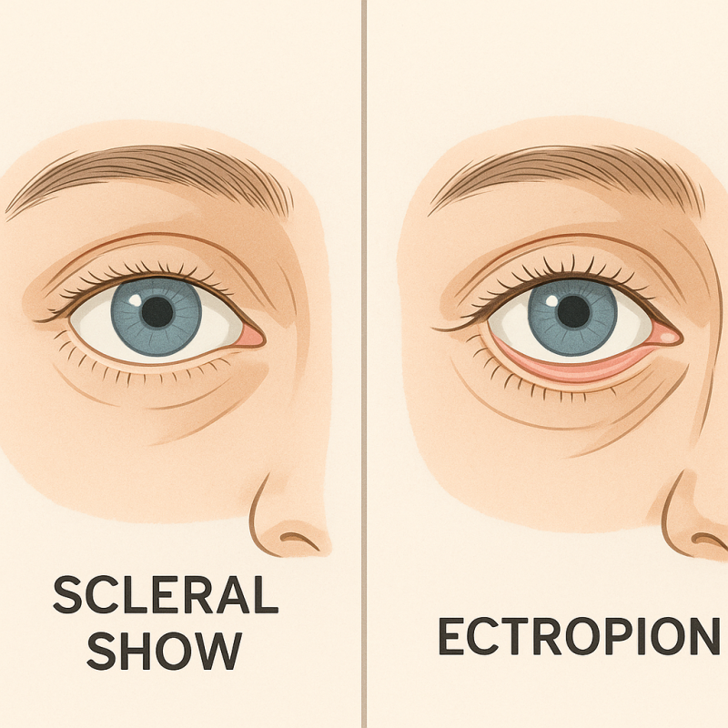
🔹 Scleral showは、下まぶたが少し下がって白目が見え、美容的に好ましくない状態
🔹 Ectropionは、下まぶたが完全に裏返って機能に問題を引き起こす病的な状態
MATERIALS AND METHODS
Clinical Assessment and Patient Selection
Between January 2016 and January 2020,
bilateral lower lid blepharoplasty on 280 patients at Humanitas Research Hospital (Rozzano, Milano, Italy)
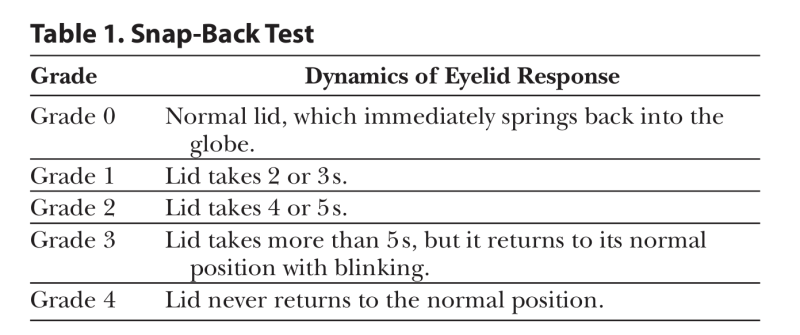
All patients were evaluated preoperatively in terms of ocular surface parameters: (a) lower eyelid retraction using “scleral show,” defined as the visible portion of the sclera between the upper rim of the lower eyelid and the inferior limbus; (b) lower eyelid laxity using the snap-back test (Table 1), the distraction test, and the assessment of eyelid margin malposition relative to the globe9; (c) amount of skin excess; (d) asymmetry; and (e) presence/ absence of orbital fat herniation (7.0.1. Adobe Photoshop Inc, California, was used to measure the distance).
Routine complete blood count and ECG were per formed in all patients. Preoperative standard photographs were collected, with the camera at a fixed 1-meter distance. Blinded-fashion preoperative and 10-month postoperative photograph analysis was performed by one independent plastic surgeon and one oculoplastic surgeon who did not execute the procedures to estimate both functional and aesthetic outcomes. Hence, postoperative photographs were scored from 1 (unsatisfactory outcome) to 5 (opti mal outcome).
Finally, at 10-month follow-up visit, a VAS from 0 to 10 was administered to all patients to assess patient sat isfaction in terms of lower lid appearance and symmetry compared to baseline. The VAS was shown to be less vul nerable to bias from confounding factors compared to other scales.10
Takeaways Question: How to prevent surgical complications in lower eyelid blepharoplasty? Findings: We highlight novel insights into a surgical technique for lower eyelid reconstructive surgery based on our 20-year experience in this specific field, which avoids the use of muscle flaps, thus preventing tarsal liga ment dystopia, limiting thermal spread, and preserving orbicularis-muscle innervation. We discuss clinical mani festations, such as epiphora, dry eye, blurred vision, eye discomfort, and photophobia, and their relation to lower lid displacement.
Meaning: Our technique helped us in preventing surgical complications, specifically lateral canthal dystopia, which ranges from round eye to ectropion. This improves post operative outcomes, revealing high patient and surgeon satisfaction over time.
A longstanding and satisfying experience with this technique let our group develop peculiar shrewdness crucial in the prevention of round eye. Particularly, our expertise focuses on the following strategical aspects:
1. The correction of horizontal skin laxity trough fixation of the lateral portion of the orbicularis oculi. Two possible surgical approaches, with or without the use of muscle flaps, can be used:
Reidy-Adamson technique: a triangular flap is harvested in the upper lateral portion of the lower orbicularis-oculi muscle. This is passed beneath a superolateral canthal tunnel and then fixed at the periosteum of the upper lateral orbital rim (above the Whitnall tubercle) to obtain optimal lower eyelid contour.
Our alternative technique, which does not imply the use of a muscle flap: during the skin excision, the orbicularis-oculi muscle is removed in a complementary fashion. Skin stitches are positioned to grasp skin-muscle-skin in an oblique fashion. This lateral vector puts into tension the muscle, compensating the horizontal skin laxity. As this approach has appeared to be very effective in a relatively short operative time, it has been used as our routine technique for more than 20 years now.
2. Sparing of tarsal ligaments, through a transcutaneous lower lid incision made 1 mm below the eyelashes line, directed to the prevention of dystopia following the tarsal ligament healing process and subsequent ectropion. In fact, misposition of tarsal ligament may lead to palpebral and bulbar conjunctiva detachment, altering the lateral final aesthetic profile of the patient.
3. Preserve the orbicularis innervation from the facial nerve by limiting inferolateral skin dissection within the orbital rim.11
4. Control of thermal spread by using cold blade dissection and diathermocoagulation in bipolar mode. We always avoid the use of cautery in monopolar mode, by preferring cold blade dissection that has been shown to be less traumatic and by avoiding histologic alteration of tissues, thus leading to less risk of conjunctival thermal injury. In case of punctate bleeding, coagulation is obtained by using bipolar forceps, which is more effective in limiting excessive thermal spread and reducing the risk of unintended injury and edema of collateral tissues, thus favoring and speeding up the healing process.
Complication rate was assessed during follow-up, made at 1 week, 2 months, and 10 months postoperatively. Specifically, patients were screened for lower eyelid malposition (including round eye, ectropion, and entropion), epiphora, dry eye, red eye, “foreign body sensation,” blurred vision, photophobia, and eye discomfort. Furthermore, the presence of infections, peribulbar hematoma/seroma, suture abscesses/cysts/granulomas, bad scarring and webbing, injury of inferior-oblique muscle or bulb, and relapse of the orbital fat were evaluated (Fig. 1A–C).
著者らの下眼瞼手術の核心戦略4つ
目標:「丸い目」(round eye、目が完全に閉じない症状)の防止+自然な目元の維持
1️⃣ horizontal skin laxityの矯正方法
・Reidy-Adamson技法:
o 下眼輪筋から三角形の筋弁(flap)を採取し、
o 目尻の上側の骨(上眼窩縁)に固定 → 目の下側の輪郭を引き上げる。
・主に用いる著者らの技法:
o 筋弁なしで皮膚を切除する際に、下眼輪筋も補完的に除去。
o 皮膚縫合時に「皮膚-筋肉-皮膚」を斜めに引っ張る → 筋肉が引っ張られ、たるんだ皮膚を引き締める。
→ 20年以上使用している、より迅速で効果的な方法
2️⃣ 眼瞼支持組織の保護
・切開位置:まつげのすぐ下(1mm) → 眼瞼を支える靭帯(tarsal ligament)の損傷防止。
o 靭帯損傷時、眼瞼が裏返ったり(外反症)、目の形が歪む可能性がある。
3️⃣ 神経の温存
・皮膚剥離時、眼窩縁の内側のみに限定的に
o 顔面神経が眼輪筋に接続する部分を保護。
4️⃣ 熱損傷の防止
・「冷却メス」のみで組織分離 → 熱による組織変性を最小限に抑える。
・出血時、両極電気凝固器(bipolar)のみ使用 →
o 熱が周辺組織(結膜など)に広がるのを遮断。
📊 安全性検証
・術後1週間/2ヶ月/10ヶ月の追跡観察で以下の合併症を検査:
o 眼瞼変形(丸い目、外反症、内反症)、感染、涙/乾燥症、視界のぼやけ、出血など。
よくある質問
下眼瞼はなぜ老化に弱いのですか?
はい、下眼瞼は皮膚の弾力低下、靭帯支持力の喪失、眼輪筋のたるみ、眼窩隔膜が薄くなることによる眼窩脂肪の突出など、様々な老化現象に非常に脆弱です。これらの変化が目の下の美容的、機能的問題を引き起こす可能性があります。
Scleral ShowとEctropionは何が違うのですか?
Scleral Showは、下まぶたが少し下がって白目が大きく見える美容的な状態を指します。一方、Ectropionは、下まぶたが完全に裏返って結膜が外部に露出し、機能的な問題を引き起こす可能性のある病理的状態です。
下眼瞼形成術後に丸い目を予防する方法は何ですか?
はい、丸い目の予防のためには、眼輪筋の外側部分を固定して水平方向の皮膚のたるみを矯正し、まつげのラインから1mm下に切開して靭帯の損傷を最小限に抑えることが重要です。また、顔面神経の眼輪筋支配を温存し、熱損傷を制御する技術を使用します。
手術時の熱損傷制御はどのように行われますか?
はい、手術時の熱損傷制御は、冷たいメスを用いた剥離と、両極性モードの電気凝固によって行われます。単極性焼灼器の使用を避け、組織損傷を減らし、結膜の熱損傷リスクを低減する方法で進められます。
眼輪筋を用いた手術技法にはどのようなものがありますか?
はい、眼輪筋を用いた手術技法には、Reidy-Adamson技法と本文で紹介されている代替技法があります。Reidy-Adamson技法は三角皮弁を使用し、代替技法は筋皮弁なしで皮膚切除時に眼輪筋を補完的に除去し、斜めに縫合して水平方向の皮膚のたるみを矯正します。
下眼瞼形成術前にどのような評価を受けますか?
はい、手術前には眼球表面パラメータの評価、下眼瞼の弛緩度検査(スナップバックテストなど)、皮膚の余剰量、非対称性の有無、眼窩脂肪ヘルニアの有無などを総合的に評価します。これにより、患者に最適化された手術計画を策定します。